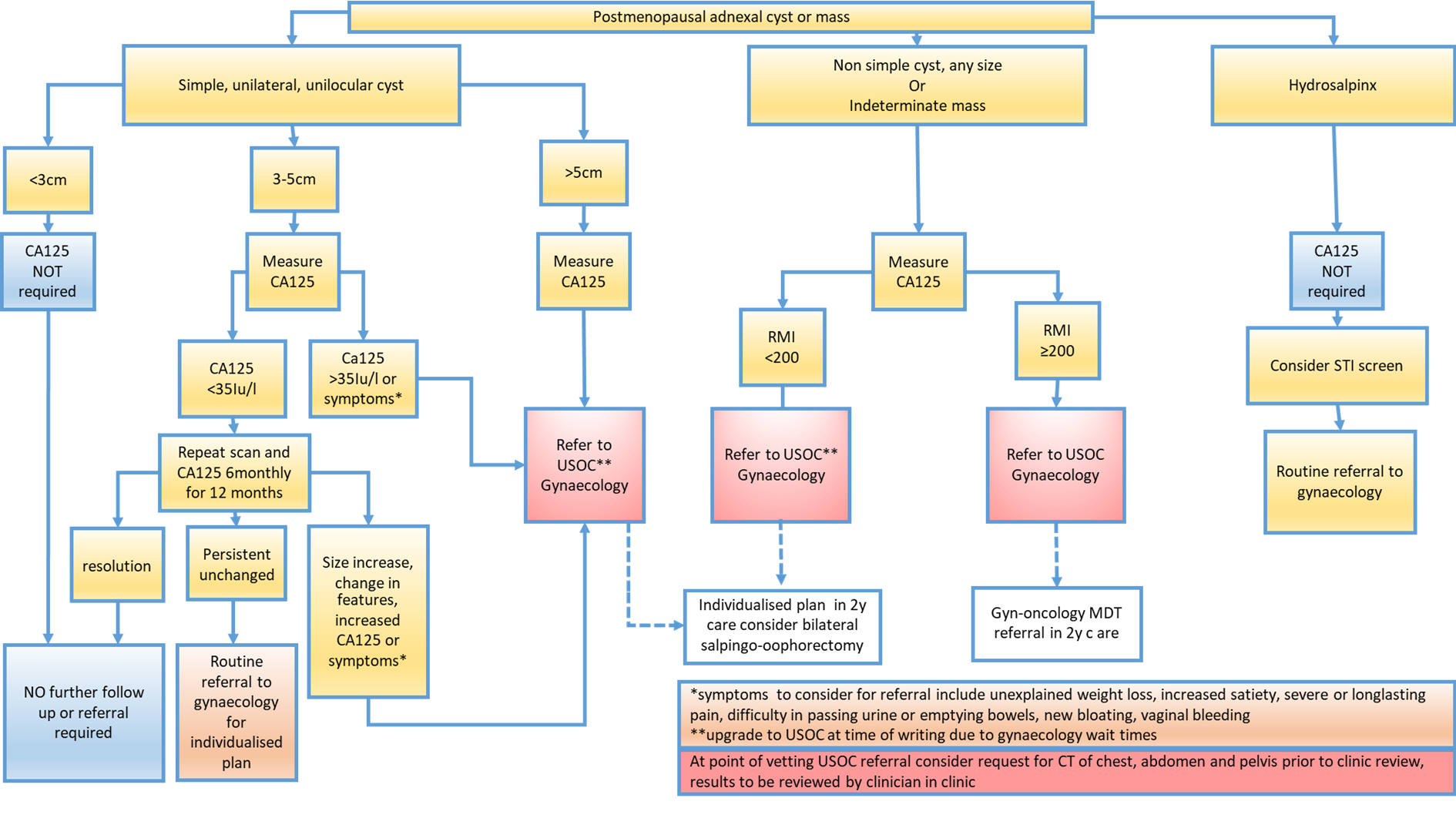
The use of RMI I scoring has been shown to be an effective method of determining women who are at low or high risk of malignancy. This will determine the need for onward referral and management required.
Use of the original RMI I calculation remains the most utilised, widely available and validated effective scoring system, with modifications using RMI II, RMI III and RMI IV systems showing no clinical benefit [2].
RMI I scoring includes measurement of CA125 and the assessment of specific ultrasound features. Therefore, ultrasound reporting must detail the morphological features present to enable calculation of the RMI I accurately.
Calculation of RMI I
The method for calculation of RMI is outlined below. The parameters used are Ultrasound score (U), Menopausal status (M) and CA125 (iu/ml).
- The ultrasound result is scored 1 point for each of the following characteristics: multilocular cysts, solid areas, metastases, ascites and bilateral lesions. U = 0 (for an ultrasound score of 0), U = 1 (for an ultrasound score of 1), U = 3 (for an ultrasound score of 2–5).
- The menopausal status is scored as 1 = premenopausal and 3 = postmenopausal. This guideline is directed at postmenopausal women and therefore all will be allocated the same score of 3 for menopausal status.
- Serum CA125 is measured in iu/ml
Interpretation of RMI I score
An RMI I of <200 is used as a cut off for malignancy risk.
If the RMI is >200, a USOC referral must be made to gynaecology and the case should then be referred to the Gynaecology/Oncology MDT via Teams for decision regarding diagnosis and further management.
A CT scan of chest, abdomen and pelvis (with contrast where appropriate) should be arranged in the interim as per West of Scotland Cancer Network, Guidelines for imaging of Gynaecological Malignancy (651). Currently, this can only be requested in secondary care and should be considered at point of vetting. If ordered at point of vetting, the results should be reviewed by the clinician who then assumes care after clinic appointment.
If the RMI is <200, but the woman is symptomatic or CA125 >35, then surgical management should be considered. Surgery should also be considered if the RMI is < 200 but the cyst >5cm in diameter.
Ovarian torsion should be managed as an emergency, and surgery in these cases should not be delayed to await gynaecological oncology team MDT discussion.