Vascular trauma associated with fractures/dislocations

Warning

Fast and accurate diagnosis is paramount if limb salvage is to be successful. Early orthopaedic and vascular consultant referral and intervention is usually indicated.

The British Orthopaedic Association have published guidelines on the mangement of vascular injuries.

Vascular injury in the setting of a fracture or dislocation can rapidly threaten limb viability, and, in exsanguinating cases, life itself. Priorities are simultaneous management of life-threatening and limb-threatening injuries, with emphasis on early recognition, haemorrhage control and restoration of perfusion.   

Management in the ED

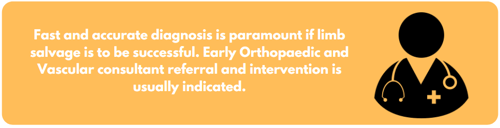
  • Treat life threatening injuries in parallel conjunction with limb threatening injuries.
  • Activate the Trauma Major Haemorrhage protocol if the patient meets criteria..
  • Control active haemorrhage
    • Direct pressure and elevation as first line
    • Consider haemostatic dressings.
    • Apply a tourniquet (placed as distal as possible) if bleeding is not controlled. Record application time.
    • Consider Foley Catheter balloon tamponade for junctional wounds.
  • Avoid blind clamping
  • Document neurovascular status (pulses, cap refill, sensation and motor function) at first contact.
  • Realign and splint the pulseless, deformed limb immediately. Then re-check and document perfusion.
  • Note: Pinkness, capillary refill or Doppler signal alone do not exclude significant vascular injury.
  • Identify direct/indirect signs of injury. 


Direct signs

Indirect signs

i.


ii.


iii.


iv.


haemorrhage


haematoma


palpable pulse


thrill/bruit

Pulsatile


Expanding


Absent


Palpable

i.

ii.

iii.

iv.

v.


vi.

Reduced or unequal pulse(s)

Non-pulsatile haematoma

History of significant haemorrhage

Injury in close proximity to neurovascular structures

Mechanism e.g. knee dislocation/displaced tibial plateau, groin contusion from handlebar or mangled extremity

Paraesthesia

Do not use ‘pinkness’, capillary return or Doppler signal to exclude injury

Direct Signs

  • Urgent surgical exploration is indicated.
  • Angiography should not delay revascularisation but may be considered if:
    • Multilevel injury.
    • Peripheral vascular disease.
    • Absent proximal pulse.
    • Patient undergoing a WBCT for other indications and scan extended to include limb angiography.

Indirect Signs

  • CT Angiography should be performed as soon as possible.

Imaging

  • Plain XR’s of the fracture/dislocation site to assess bony alignment
  • CTA from proximal to distal vessel segments if
    • Hard signs and patient is stable enough for a quick scan
    • Any soft signs
    • Shotgun, high-velocity injuries, or complex fracture patterns
  • Do not let imaging delay revascularisation if the limb is ischaemic and the patient is unstable

Surgical priorities and combined ortho-vascular approach

  • Orthopaedic and vascular consultant should attend.  Plastic surgery should attend if open fracture or major soft tissue loss.
  • Patients should be aware of the high risk of amputation for some injuries.
  • Two consultants should be involved in the decision to perform early amputation.
  • Beyond 3-4 hours, warm ischaemia results in irreversible tissue damage and an increasing risk of amputation.  Risks of delayed revascularisation include myogobinuria and may be associated with increased mortality.  Access incisions should be planned to facilitate soft tissue coverage.
  • Revascularisation:
    • Vascular repair usually precedes definitive fixation of the fracture (unless external fixation for quick stabilisation is needed first to facilitate vascular repair)
    • Temporary vascular shunts may be inserted to restore flow before definitive repair if the limb is threatened or in a damage control scenario
  • Aim to restore flow within 3-4 hours of injury to minimise muscle necrosis and reduce amputation risk
  • Prioritise haemorrhage control and revascularisation.  This may involve external fixation and temporary shunts.

Intraoperative Considerations

  • Plan incisions with future soft tissue coverage in mind
  • Fasciotomies:
    • Low threshold, especially after prolonged ischaemia or massive transfusion
    • Decompress all compartments at risk
  • Nerve repair is typically delayed unless there is an open, clearly transacted nerve amenable to direct suture at time of vascular repair and the patients physiology allows.

Post-operative Care

  • Admit all patients to a critical care environment post operatively. MTW for some patients. 
  • Monitor pulses, cap refill, motor/sensation every 1-2 hours initially
  • Antibiotics as per local guidelines
  • DVT prophylaxis unless actively bleeding or coagulopathic
  • Early physiotherapy to maximise limb function

Editorial Information

Last reviewed: 01/04/2025

Next review date: 01/04/2028

Version: 1.0